how do they x ray babies hips
After around 4 to 6 months of age X-rays are the preferred method for evaluating and monitoring hip dysplasia. It has nothing to do with whether or not the parents will hold the infant.

How To Position The False Profile View X Ray Of The Hip X Ray Profile View Positivity
Hip X-rays are done with a child lying on a table.

. At birth the baby cant move the thigh outward at the hip as far as normally possible. The purpose of this study is to report US results and follow-up of. Hip problems may not be present at birth.
They are of more concern. X-rays can be taken once your baby is 3 months old. Subsequent x-rays will track the hip joints progress.
Computed tomography scanogram for leg length discrepancy assessment. Thats because most of a babys hip joint is still soft cartilage which wont show up on an X-ray. I was also shown an X-ray that suggests he will have hip joint issues lending towards arthritis.
If a physical exam an ultrasound or an X-ray confirm a diagnosis your pediatrician will likely refer you to a pediatric orthopedic specialist for continued care and treatment. Hip ultrasounds take less than 20 minutes and the child will not feel any pain during the examination. It occurs more commonly in boys typically between 5 and 8 years of age but may range from the ages 3-12.
This helps to see blood vessels and blood flow on the X-ray. After around 4 to 6 months of age X-rays are the preferred method for evaluating and monitoring hip dysplasia. A hip click can be felt by the examiner when the hip joints may not have formed normally.
Computed bone maturity bone age measurement are performed in cases of suspected growth delay or early pubertal development. They give your healthcare provider information about structures inside the body. It is the preferred way to diagnose hip dysplasia in babies up to 6 months of age.
Because of the risk of developmental dysplasia of the hip in infants born breech-despite a normal physical exam-the American Academy of Pediatrics AAP guidelines recommend ultrasound US hip imaging at 6 weeks of age for breech females and optional imaging for breech males. Perthes disease also known as Legg-Calvé-Perthes disease is an idiopathic avascular necrosis of the proximal femoral epiphysis. X-rays have more energy than rays of visible light or radio waves.
An X-ray technician will take pictures of the hip. The doctor first checks your babys hips in the hospital after birth. An AP radiograph should be obtained with hips in the neutral position.
Then a surgeon gently pushes the ball of their thighbone joint into the hip socket where it belongs. However x-rays of the mothers lower torso - abdomen stomach pelvis lower back or kidneys - may expose the unborn child to the direct x-ray beam. Your baby was born in the breech position after 28 weeks of.
If they are obtained in the newborn period the hips should be placed at 20-30 A diagnosis on X-ray is made after Hilgeureiners H. F ratio 18 firstborn baby. X-rays are forms of radiant energy like light or radio waves.
A hip ultrasound might be done for a baby if the doctor finds a hip problem such as. Inflammation where your sacrum joins. Most children do not need surgery but for those who do an arthrogram x-ray dye injected into the hip joint at the beginning of the surgery can help the surgeon decide exactly what needs to be corrected.
They can penetrate your body. Its a cast that goes around both hips and down the leg to keep the hips aligned. A pelvic X-ray can help your doctor detect various conditions such as.
If she does have it they may try to brace it first. You will go in the room with him he will need to be stripped from the waist down they will take x-rays of him flat on his back legs dead straight and together you wil be able to hold him in this position then an x-ray of his still on his back with his knees bent facing outwards and the soles of his feet put together he will be fine. Computed bone maturity bone age assessment.
Because they spin around the body taking multiple images CT scans can deliver radiation doses that are up to 200 times higher than an. In addition exposing the parents to ionizing radiation X-rays needlessly goes against the ALARA principle. The reported incidence of developmental dysplasia of the hip varies between 15 and 20 per 1000 births 1 with the majority 60-80 of abnormal hips resolving spontaneously within 2-8 weeks 1 so-called immature hip.
During treatment x-rays can reveal the progress of the hip as it improves. Risk factors include 14. An ultrasound may be needed to get a picture of the hip.
These tests expose children to low doses of radiation. Two tests are performed called the Barlow and Ortolani tests to examine the function of the hip joints. About one in eight scans ordered for kids is a CT scan.
Arthritis that affects your hip. But for babies with an abnormal physical exam or major risk factors for developmental dysplasia of the hip or DDH family history Breech position etc the AAP supports referral for. It can occur bilaterally but it is usually asymmetric.
X-rays can be taken once your baby is 3 months old. In babies with hip dysplasia the joint has not formed normally and the hips are prone to moving in and out of joint. The American Academy of Pediatrics does not recommend routine ultrasounds for every infant.
Hip ultrasounds are a safe non-invasive procedure that does not use any radiation. It is put on by an orthopedic surgeon while using. X-rays are a kind of imaging test.
Vertical lines have been drawn and the proximal femoral epiphysis is seen to have lateral or proximal displacement indicating. They do this by gently pushing and pulling the babys thigh bones to see if they are loose in the hip socket. Horizontal and Perkins P.
If it persists they may put on a spica cast. The doctor hears or feels a hip click when moving the infants thigh outward. How is hip dysplasia treated in babies.
Ultrasounds use inaudible sound waves which bounce off of the bones and muscles to create an image for radiologists to interpret. Pregnancy is a time to take good care of yourself and your unborn child. Appointments and Referrals.
What Are the. From the front anteroposterior view or AP from the side lateral view also known as the frog leg lateral view Typically X-rays of both hips are taken for comparison even if only one hip is causing symptoms.

How To Shower After Hip Replacement Surgery Livestrong Com Hip Replacement Surgery Hip Replacement Exercises Hip Brace

Causes Of Ddh Hip Dysplasia Baby Developmental Dysplasia Of The Hip Baby Wearing

Hip Joint Developmental Hip Dysplasia 1 Year Old Child With A Dislocated Right Hip The Degree O Developmental Dysplasia Of The Hip Radiography Subluxation

Pin By Meg Carter On Ortho Hip Dysplasia X Ray Orthopedics

Developmental Dysplasia Of The Hip Ddh Diagnostic Imaging Developmental Dysplasia Of The Hip Diagnostic Imaging Case Study

Lower Limb Radiographs Anatomy And Physiology Anatomy Sacroiliac Joint
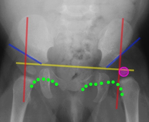
Lines Of The Hip Pediatrics Pediatrics Pediatric Nurse Practitioner Pediatric Radiology

X Ray Image Of Child Swallowed The Coins For A Medical Diagnosis Medicine Pictures Children Images X Ray Images

Hip Dysplasia In Adolescents And Young Adults Hss Hipproblems Hip Dysplasia Hip Problems Hips

Congenital Hip Dislocation Chd Happens When A Child Is Born With An Unstable Hip Read On To Learn More Ab Canine Hip Dysplasia Hip Dysplasia Hip Dislocation

Uk Professor Says Swaddling Epidemic Gives Babies Clicky Hips Daily Mail Online Hips Professor Baby Swaddle

Pin On X Rays
Periacetabular Osteotomy X Rays X Ray Ehlers Danlos Syndrome Writing A Book

Radiology Radiologic Imaging Signs List Collection Illustrated Cases Xray X Ray Gi Gu Chest Th Radiology Avascular Necrosis Avascular Necrosis Hip

Diagnosis Prevention And Management Of Canine Hip Dysplasia A Revie Vmrr Canine Hip Dysplasia Diagnostic Imaging Total Hip Replacement

Hip Dysplasia When You Re Too Young For A Hip Replacement Periacetabular Osteotomy Pao Bursitis Hip Hip Replacement Hip Replacement Surgery

One Month Post Op From A Right Periacetabular Osteotomy To Correct Hip Dysplasia Ehlers Danlos Syndrome Hip Dysplasia Surgery Recovery

Pin On Nursing 1st Semester

Pin On Fibro Autoimmune Diseases